Showing posts with label bone. Show all posts

Saturday, May 21, 2022

Electrical Bone Growth Stimulator

A bone growth stimulator is a supplemental device worn following cervical neck or lumbar low back spine surgery. Global Market insights 2017 to 2022.

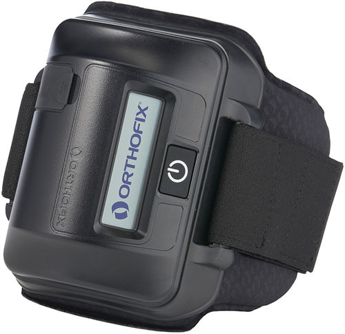
Orthofix Announces Nass Coverage Policy Recommendations For Electrical Bone Growth Stimulators Covering The Specialized Field Of Orthopedic Product Development And Manufacturing

An electric bone growth stimulator uses electric current to promote bone healing.

Electrical bone growth stimulator. Ad Shop Devices Apparel Books Music More. Electrical stimulation works by promoting the bodys healing response after a fracture has occurred. Electrical Bone Growth Stimulators Osteogenic Stimulation.

5 rows Electrical Bone Growth Stimulation. Description The new OsteoGen Dual Lead Stimulator offers 40 microamperes in two leads each with 20 microamperes. A bone growth stimulator also known as a bone stimulator is a durable medical equipment designed to deliver electric current or ultrasound waves to the bone in order to stimulate the osteoblastic function and trigger the growth of new cells.

When bone is broken or bent it. Electrical or electromagnetic bone growth stimulators may be indicated when ALL of the following are present. Available in both Mesh and Straight cathodes the OsteoGen Dual Lead Stimulator provides superior flexibility with any of your bone graft techniques when managing high risk fractures.

Electrical bone growth stimulation is a medical technique to promote bone growth in difficult to heal fractures by applying a low electrical current to the fracture site. 5 rows Electrical Bone Growth Stimulation. Electrical stimulation is a technique that has been used to try to accelerate bone healing.

Factmr May 17 2021. Electrical Bone Growth Stimulator Market Business Opportunities Survey Growth Analysis And Industry Outlook Bone Growth Stimulator Market Forecast Trend Analysis Competition Tracking. Non-invasive semi-invasive and invasive methods of electrical bone growth stimulation are available.

The devices stimulate the natural healing process of bone by sending lowlevel pulses of electromagnetic energy to the injury or fusion site. The use of a device either implanted into the body or. An electrical osteogenesis stimulator is a device that provides electrical stimulation to augment bone repair.

In this way it can be quite. Studies have shown that the rate of nonunion of a fracture does seem to be reduced in people who have used electrical stimulation. Bone growth stimulation is a technique of promoting bone growth in difficult to heal fractures.

The use of a device either implanted into the body or. Two types of bone growth stimulators currently exist. Spinal hcpcs codes e0748 e0749 An invasive or noninvasive spinal electrical bone growth stimulator as an adjunct to lumbar spinal fusion surgery is considered medically necessary for ANY of the following indications associated with.

BGS may be utilized to help spinal bone fuse after a fusion procedure or as a treatment for failed fusion. Electrical bone growth stimulator. Free UK Delivery on Eligible Orders.

To help overcome these healing challenges doctors commonly prescribe a treatment called bone growth therapy. Bone growth stimulation BGS is a therapy your surgeon may prescribe following a spinal fusion procedure. A variety of invasive and noninvasive interventions are used to treat fracture nonunion including im- mobilization casting open.

Bone growth stimulator is being used as adjunctive treatment to lumbar spine fusion. Bone growth stimulation is utilized to promote bone healing in difficult to heal fractures or fusions by applying electrical or ultrasonic current to the fracturefusion site.

Friday, May 21, 2021

Bone Density Study Guide

-in vertebrae and ends of long bones. Coverage of common and unique positioning procedures includes special chapters on trauma surgical radiography geriatricspediatrics and bone.

Bone Density Test Osteoporosis Screening T Score Interpretation

-less dense softer weaker.

Bone density study guide. Its important for applicants to understand the whys of what they do as well as how and what so the certification process is not just book learning Haselhuhn says. Medicine study guide bone densitometry pdf arrt bone density study guide - free ebooks. Hips pelvis spine including vertebral fracture assessment.

Bone densitometry Registry Review Exam Books ARRT study guide courses schools anatomy practice practice test Bonedensitytutor - Bone density Training Online - Scan Analysis Study Guide support. Bone densitometry Registry Review Exam Books ARRT study guide courses schools anatomy practice practice test Bonedensitytutor - Bone density Training Online - Study Guides support. DXA bone density study 1 or more sites appendicular skeleton peripheral eg.

Arrt Bone Density Study Guide Author. Arrt bone density study. BoneDensityTutor is a 21st Century study tool to help you become certified.

These materials contain recommendations from experienced RTs for bone densitometry registry review. The following study guides and resources are available to help you prepare for the bone densitometry certification exam offered by the American Registry of Radiologic Technologists. Computed tomography bone mineral density study 1 or more sites.

Arrt Bone Density Study Guide Keywords. These specs which come as part of the application handbook spell out the content areas covered in the ARRTs certification exam. Axial skeleton eg hips pelvis spine OfficeFreestanding Global 322 11621 Facility Professional 035 1263 76977 Ultrasound bone density measurement and interpretation peripheral sites any method OfficeFreestanding Global 020 722 Facility Professional 008 289.

Trabecular has a higher surface area good area for obtaining Ca. Radius wrist heel Professional -26 1113 Technical -TC 1723 5521 5986 Global 2835 77085 Dual-energy X-ray absorptiometry DXA bone density study 1 or more sites axial skeleton eg. You can have a full-time job just doing DEXA.

-highly vascular contains red bone marrow where hematopoiesis occurs. Compact bone hard outer surface. Use the bone densitometry examinations as a study guide.

Does anyone have any tips with studying for the Bone Densitometry registry. Many technologists add Bone Densitometry or DEXA scanning registry to their list of accomplishments. The Bone Density Registry is another certification offered by the ARRT.

Density study guide documents study guides for radiologic technology registry bone densitometry for technologists. DEXA is considered a separate modality in the radiology department. Online Library Arrt Bone Density Study Guide Comprehensive full-color coverage of anatomy and positioning makes Merrills Atlas the most in-depth text and reference available for radiography students and practitioners.

Bone density study guide Length. DXA bone density study 1 or more sites appendicular skeleton peripheral eg radius wrist heel Professional -26 1045 Technical -TC 2343 5521 6230 Global 3388 77085 Dual-energy X-ray absorptiometry DXA bone density study 1 or more sites axial skeleton eg hips pelvis spine including vertebral fracture assessment. Bone densitometry Registry Review Exam Books ARRT study guide courses schools anatomy practice practice test Bonedensitytutor - Bone Density Training Online - Basic Concepts Part 1 - Study Guide support.

Please remember that the demo is only a small sample of what is available once you gain full access. Bone density study guide Author. Several interactive study tools and guides are available along with many practice quizzes covering each of the bone density specific subjects to help you prepare without causing you to be overwhelmed.

Features Fullscreen sharing. BoneDensityTutor strives to go beyond the textbook and offer up to date content.

Friday, May 14, 2021

Bone Density Test Guidelines

Reported as a T-score Osteopenia As above with T-score between -10 SD and -25 SD Fragility fracture. The test can identify osteoporosis determine your risk for fractures broken bones and measure your response to osteoporosis treatment.

Bone Density Testing In Clinical Practice

Use either FRAX without a bone mineral density BMD value if a dual-energy X-ray absorptiometry DXA scan has not previously been undertaken or QFracture within their allowed age ranges to estimate 10-year predicted absolute fracture risk when assessing risk of fracture.

Bone density test guidelines. A bone mineral density BMD of 25 standard deviations SD or more below the mean peak mass of average of young healthy women as measured by dual-energy X-ray absorptiometry DXA. This is shortened to DEXA. However longer intervals may be adequate for repeated screening to identify new cases of osteoporosis.

The logic behind extending testing intervals. Youre a woman whose doctor determines youre estrogen deficient and at risk for osteoporosis based on your medical history and other findings. Several types of bone density tests exist.

Total body bone density scans can also be used to measure the amount of bone fat and muscle in the body. The Taskforce considers these testing frequencies to be clinically appropriate. Some radiation is absorbed by the bone and soft tissue and some travels through your body.

The preferred test is now the bone density mineral test. These guidelines recommend that testing of bone mineral density be performed based on the patients risk factors and is not indicated unless the results will affect a treatment or regimen program. Your bone density measurements will be compared with the bone density.

During a bone density scan a special type of X-ray called dual energy X-ray absorptiometry is passed through your body. Patients with a bone mineral density t-score of less than -15 and above -25 will be eligible for one scan every two years item 12322. Theyre often used to help diagnose bone-related health problems such as osteoporosis or to assess the risk of getting them.

One T-score unit eg. The most commonly used BMD test is called a central dual-energy x-ray absorptiometry or central DXA test. The test called a DEXA scan is a kind of X-ray.

Because of limitations in the precision of DEXA testing a minimum of 2 years may be needed to reliably measure a change in bone density. Many people get a bone-density test every few years. In women 65 years of age and older at baseline screening with a T-score of -150 to -199 at any site and with no.

Bone density should also be measured in women between the ages of 50 and 65 who are considered high risk for osteoporosis. In addition to obtaining your initial DEXA scan results repeating the scan every two to five years is recommended depending on. If initial bone density testing shows you have a T-score of -200 to -249 at any site or if you take medications.

Patients with a bone mineral density t-score of -15 or above will be eligible for one scan every 5 years item 12320. While it is a quick and unobtrusive procedure there are some things you should do to prepare yourself for it. New guidelines released Thursday by the National Osteoporosis Foundation NOF call for bone mineral density BMD testing for all men ages 70 and older and for younger men with risk factors for.

A bone-density test is a way to measure the strength of your bones. Special detectors in the DEXA machine measure how much radiation passes through your bones and this information is sent to a computer. Medicare Part B Medical Insurance covers this test once every 24 months or more often if medically necessary if you meet one of more of these conditions.

The main reason to have the test is to find and treat serious bone loss called osteoporosis and prevent fractures and disability. Bone density scans also known as DEXA scans help to work out your risk of breaking a bone. A bone mineral density BMD test is can provide a snapshot of your bone health.

To diagnose osteoporosis doctors will usually use a reliable means of measuring a patients bone density by performing a procedure known as dual energy X-ray absorptiometry DEXA. -10 is roughly 10 of ones. Read on to learn how to prepare for a bone density scan.

Medicare removed other similar tests when.

Tuesday, April 28, 2020

Magnetic Bone Stimulator

When the initial results were analyzed 10 additional study patients were added. Orthofix Bone Growth Therapy devices provide a safe noninvasive treatment that helps promote healing in fractured bones and spinal fusions that have not healed or have difficulty healing.

A Closer Look At Bone Stimulators For Charcot Podiatry Today

The mechanism of osteogenesis is not clear therefore studies look for empirical evidence.

Magnetic bone stimulator. 11 The demineralized bone matrixinduced EO model developed by Hari Reddi has been extremely useful for examining the details of. Electrical bone growth stimulators are a supplemental form of therapy to help enhance the bodys bone healing process a process that is absolutely essential for the success of any type of spinal fusion surgery. BGS may be utilized to help spinal bone fuse after a fusion procedure or as a treatment for failed fusion.

The effectiveness of electrical stimulation and Pulsed Electro Magnetic Field PEMF stimulation for enhancement of bone healing has been reported by many workers. CMF Spinalogic is a portable battery-powered micro-controlled noninvasive bone growth stimulator indicated as an adjunct electromagnetic treatment to primary lumbar spinal fusion surgery for one or two levels. The SpinaLogic bone growth stimulator is a nonsurgical treatment that your physician may prescribe to help the healing of your spine surgery.

Fundamental and practical aspects of therapeutic uses of pulsed electromagnetic fields PEMFs Effects of static magnetic and PEMFs on bone healing Strong static magnetic field stimulates bone formation to a definite orientation in vitro and in vivo Use of physical forces in bone healing. 68 Positive results and 30 min wear time can encourage patient compliance. Bone stimulators are not indicated for fractures of cancellous bone fracture gaps greater than 50 percent of the diameter of the bone at the level of the fracture synovial pseudarthrosis when there is fixation with magnetic materials in pregnant women or in patients with skeletal immaturity.

In a well-characterized in vivo model of EO PEMF stimulation has been shown to increase the synthesis of cartilage and bone matrix growth factors and to enhance the proliferation and differentiation of osteoblast-like primary cells. Thirty-one subjects were studied. Magnets have been used in alternative medicine for conditions ranging from systemic illness to joint pain.

Bone injuries meaning broken cracked or severed bones often have trouble healing quickly or completely due to a variety of natural causes warns Orthofix. Designed to be a lightweight device with cushion strap designed for maximum patient comfort. A bone stimulation device emits low-level electromagnetic pulses on the bone injury location promoting natural bone growth and fusion.

The devices stimulate the natural healing process of bone by sending lowlevel pulses of electromagnetic energy to the injury or fusion site. Naturally you have questions about this technology. 20 In addition one would not employ a bone stimulator in patients with.

When bone is broken or bent it generates an electrical field. One or two treatment coils placed into a brace or directly onto the skin that deliver a type of electromagnetic field called a Pulsed ElectroMagnetic Field PEMF or a Combined Magnetic Field. The idea is that magnets might improve circulation and increase blood flow and thus the delivery of nutrients to the fractured bone.

The stimulator uses a very low-strength Combined Magnetic Field CMF to activate the bodys natural healing process. Human bone is actually a living tissue and like skin has the inherent ability to heal itself when broken or injured. The SpinaLogic bone growth stimulator has been proven in clinical studies to increase to.

To help overcome these healing challenges doctors commonly prescribe a treatment called bone growth therapy. The devices stimulate the bones natural healing process by sending low-level pulses of electromagnetic energy to the injury or fusion site. Powered by CMF Bone Growth Stimulation The OL1000 and Spinalogic powered by DJOs more advanced technology 7 provides medical professionals with an effective tool for healing non union fractures and primary lumbar spinal fusion procedures.

Similarly some have proposed using magnets to help heal bone fractures. This study was designed to assess the value of combined magnetic field CMF bone growth stimulation in the treatment of acute phase 1 Charcot neuroarthopathy. A bone growth stimulator is a supplemental device worn following cervical neck or lumbar low back spine surgery.

Bone growth stimulation may be utilized to help spinal bone fuse after a. A bone stimulator delivers ultrasonic or pulsed electromagnetic waves to the nonunion site to stimulate healing. Initially 10 controls and 11 study patients were examined.

Affordable Care Act Flu Shot

Scott Breidbart MD. Under the Affordable Care Act health insurers are required to cover flu and other vaccines without charging a. Vaccin...